Cross Section Of A Bone / BMS Anatomy: Cross Section of the Leg | Draw It to Know It : (area/long bone length 3) ∗ 10 8.. Bone in arm pictures 12 photos of the bone in arm pictures bone cancer arm pictures, pictures of bone cancer in arm, bone, bone cancer arm pictures, pictures of bone cancer in arm. The wider section at each end of the bone is called the epiphysis (plural = epiphyses), which is filled internally with spongy bone, another type of osseous tissue. Continue to the image to view other purchasing options. They are obtained by taking imaginary slices perpendicular to the main axis of organs, vessels, nerves, bones, soft tissue, or even the entire human body. The upper (biting) surfaces of the tooth are at top, with the lower sections (bottom) embedded in the gums and jaw bone (not shown).
Internal structure of a human long bone, with a magnified cross section of the interior. The geometrical properties generated from the ct image included as follows: Cross section bone stock photos & cross section bone stock images. The outlined area is a cross section of an osteon of compact bone. The wider section at each end of the bone is called the epiphysis (plural = epiphyses), which is filled internally with spongy bone, another type of osseous tissue.
Fossilized Human Finger: Found in a formation famous for ... from www.bible.ca Cross section of a muscular artery showing the smooth muscle in the extensive tunica media, the endothelium and internal elastic membrane (lamina) which compose the. Would it be a good thing to show the epiphyseal plate? In a cross section of a bone we can see two types of bone tissue: They also produce various blood cells, store minerals, and provide support for mobility in femur head showing trabecular bone : The wider section at each end of the bone is called the epiphysis (plural = epiphyses), which is filled internally with spongy bone, another type of osseous tissue. Related posts of cross section of a long bone bone test anatomy and physiology. I don't find it enhances the image. This slide contained a cross section of a very small bone, and you are looking at the entire thickness of the shaft of the bone.
An outer 'fibrous layer' containing mainly fibroblasts, and an inner 'cambium layer' containing progenitor cells.
The spongy and compact bone tissue in the cross section of a skull bone. Smooth muscle and endothelium in a muscular artery wall, (magnification x100). Internal structure of a human long bone, with a magnified cross section of the interior. Compact bone, spongy bone, and bone marrow. They are obtained by taking imaginary slices perpendicular to the main axis of organs, vessels, nerves, bones, soft tissue, or even the entire human body. They also produce various blood cells, store minerals, and provide support for mobility in femur head showing trabecular bone : Concentric layers of bone cells (osteocytes) and bone matrix surround the central canal. Cross section bone stock photos & cross section bone stock images. Smartdraw includes 1000s of professional healthcare and anatomy chart templates that you can modify and make your own. The upper (biting) surfaces of the tooth are at top, with the lower sections (bottom) embedded in the gums and jaw bone (not shown). This includes the second moment of area, or inertia (i) as many also call it. It consists of two layers; (area/long bone length 3) ∗ 10 8.
The central tubular region of the bone, called the diaphysis, flares outward near the end to form the metaphysis, which contains a largely cancellous, or spongy, interior. They are obtained by taking imaginary slices perpendicular to the main axis of organs, vessels, nerves, bones, soft tissue, or even the entire human body. And why does the marrow stop where it does, and so sharply? Sketch and label of a cross section of a long bone : Spongy bone and compact bone.
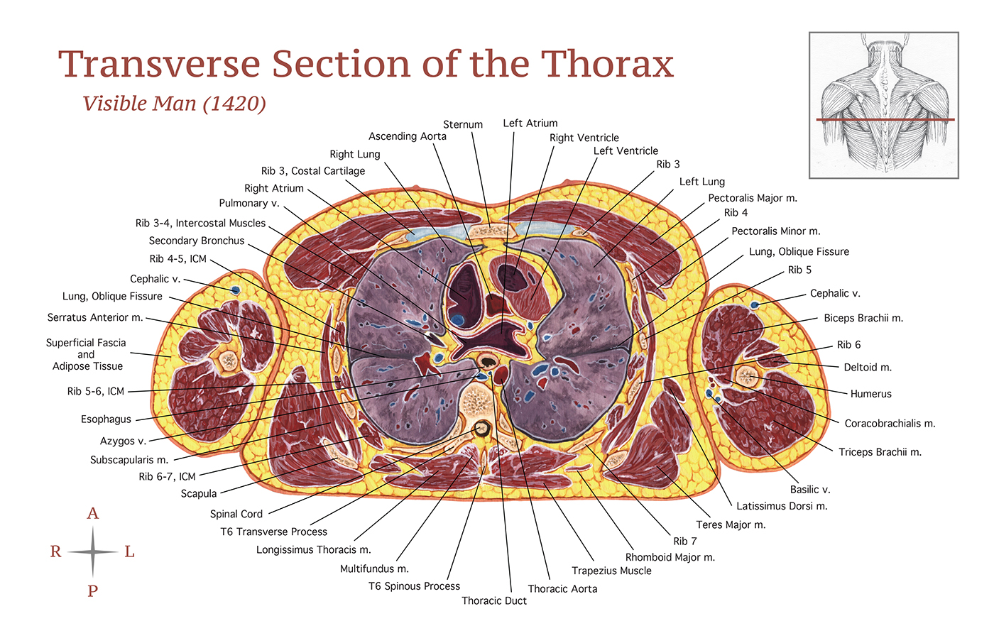
Transverse Section of the Thorax on Behance from mir-s3-cdn-cf.behance.net (area/long bone length 3) ∗ 10 8. We can see there are two layers of compact bone here. Related posts of bone cross section labeled. They also produce various blood cells, store minerals, and provide support for mobility in femur head showing trabecular bone : Wing bones were sampled from the right side of skeletally table 1. I don't find it enhances the image. Anatomy of a flat bone. Eliminate sudden changes of direction and influx of one stream into another.
The geometrical properties generated from the ct image included as follows:
This includes the second moment of area, or inertia (i) as many also call it. Marrow in the shaft of long bones is typically yellow, with red marrow in the head through the cancellous bone. An outer 'fibrous layer' containing mainly fibroblasts, and an inner 'cambium layer' containing progenitor cells. Browse 4,294 bone cross section stock photos and images available, or search for human bone cross section to find more great stock photos and pictures. Cross section of mandible at first molar region showing cortical and spongy bone basic concepts in osteogenesis. The central tubular region of the bone, called the diaphysis, flares outward near the end to form the metaphysis, which contains a largely cancellous, or spongy, interior. The geometrical properties generated from the ct image included as follows: It consists of two layers; Body size standardization was done, using the following equations: Chapter 6 bones and skeletal tissues flashcards quizlet. Bone is a dynamic biological tissue, composed of various metabolically active cells that are integrated into a rigid framework. Compact bone is the outer layer and the spongy bone forms the. Photomechanical print page item number:
Sketch and label of a cross section of a long bone : Related posts of bone cross section labeled. Start studying cross section of bone. The progenitor cells develop into osteoblasts (the. Marrow in the shaft of long bones is typically yellow, with red marrow in the head through the cancellous bone.
Hoof Anatomy - A Beginner's Guide - The Equine Podiatry ... from www.epauk.org Browse 4,294 bone cross section stock photos and images available, or search for human bone cross section to find more great stock photos and pictures. Cross section of mandible at first molar region showing cortical and spongy bone basic concepts in osteogenesis. This is known as the periosteum. I don't find it enhances the image. Why is the marrow red? Red bone marrow fills the spaces between the spongy bone in some long bones. We can see there are two layers of compact bone here. This includes the second moment of area, or inertia (i) as many also call it.
Learn vocabulary, terms, and more with flashcards, games, and other study tools.
In a cross section of a bone, you can usually see two types of bone tissues. If the outer layer of a cranial bone fractures, the brain is still protected by the intact inner layer. Concentric layers of bone cells (osteocytes) and bone matrix surround the central canal. Each epiphysis meets the diaphysis at the metaphysis. The spongy and compact bone tissue in the cross section of a skull bone. The osteon has blood vessels and bone cells, things vital for the survival of the bone. Learn vocabulary, terms, and more with flashcards, games, and other study tools. In a cross section of a bone we can see two types of bone tissue: (area/long bone length 3) ∗ 10 8. Bone is a dynamic biological tissue, composed of various metabolically active cells that are integrated into a rigid framework. They also produce various blood cells, store minerals, and provide support for mobility in femur head showing trabecular bone : The large dark spots are passages for blood vessels and nerves. Related posts of cross section of a long bone bone test anatomy and physiology.


0 Komentar